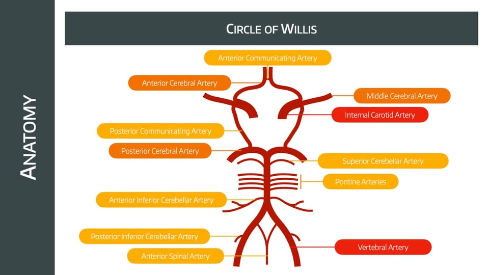
The Circle of Willis

Take home messages
- Neurons die after five minutes of no blood flow
- The Circle of Willis is a unique bilateral arterial anastomosis
- 70% of ischaemic strokes are in the anterior circulation
Weirdly popular
For a structure that you never really see or do anything to or for as an anaesthetist, the Circle of Willis gets examined extraordinarily frequently.
You need to be able to draw it, and to label it, and to answer some questions about it.
Key facts about the Circle of Willis
- It is an arterial anastomosis
- It receives around 700ml/min (50ml/100g/min of brain tissue) of blood
- That's around 15% of the total cardiac output
- 2/3rds of the supply arrives via the internal carotid arteries
- 1/3rd via the vertebral arteries
The Circle of Willis primarily exists as an insurance policy against vascular occlusion. Loss of one artery should hopefully be compensated for by the anastomotic ring, and cerebral perfusion preserved.
Which arteries form the Circle of Willis?
- Two internal carotid arteries (from the common carotids)
- Two anterior cerebral arteries
- One anterior communicating artery
- Two posterior communicating arteries
- Two posterior cerebral arteries (arising from the basilar artery)
The middle cerebral arteries, while very important, aren't technically part of the circle - they supply the lateral cerebral cortex
What does the anterior cerebral artery supply?
- Superomedial parietal lobe
- Medial frontal lobe
- Corpus callosum
- Prefrontal cortex
- Primary motor cortex (mainly lower limbs)
- Broca's area
What does the middle cerebral artery supply?
- Lateral frontal lobe
- Lateral parietal lobe
- Lateral temporal lobe
- Basal ganglia
- Internal capsule
- Motor and sensory areas to face and upper limbs
What does the posterior cerebral artery supply?
- Occipital lobe
- Medial temporal lobe
- Thalamus
- Part of the internal capsule
Where are arterial aneurysms most commonly found?
- 30-40% at junction of anterior communicating and anterior cerebral arteries
- 30% at junction of internal carotid and posterior communicating artery
- 20-30% in middle cerebral artery
- 10% in posterior circulation (basilar and superior and posterior inferior cerebellar arteries)
How much of the arterial flow does each artery receive?
- ICA 36%
- MCA 21%
- Basilar 20%
- Vertebral 15%
- ACA 12%
- PCA 8%
- Ophthalmic 2%
This is probably more than you need for the exam, so don't stress about this bit.
What factors affect flow in a blood vessel?
- Length of the vessel
- Diameter of the vessel
- Viscosity of blood
- Pressure gradient
Resistance = (8 x length x viscosity)/ π x radius4
So you can see that changing the radius is the most effective way to alter flow.
What factors affect cerebral autoregulation?
- CO2
- PaO2
- pH
- Lactate
- Potassium
What proportion of strokes occur in the anterior circulation?
- 70%
- 90% of these are in the MCA
Where is the only place in the body that an artery passes through a venous structure?
- Cavernous sinus
A reminder of the important graphs


Read our full post on cerebral blood flow and autoregulation here

Useful Tweets
The famous cerebral autoregulation curve, not as robustly proven as one might think ! https://t.co/hUtuBb5VCS
— Joel Neves Briard (@JNevesBriard) October 15, 2023
Are We Ready for Clinical Therapy based on Cerebral Autoregulation? A Pro-con Debate
— Critical Care Reviews (@CritCareReviews) May 14, 2023
CCR Journal Watchhttps://t.co/Sp06oA6IDG pic.twitter.com/0ZrV1RKP6Y
References and Further Reading


Primary FRCA Toolkit
Members receive 60% discount off the FRCA Primary Toolkit. If you have previously purchased a toolkit at full price, please email anaestheasier@gmail.com for a retrospective discount.

Discount is applied as 6 months free membership - please don't hesitate to email Anaestheasier@gmail.com if you have any questions!
Just a quick reminder that all information posted on Anaestheasier.com is for educational purposes only, and it does not constitute medical or clinical advice.
