Calcium channel blockers

If you're in revision mode, then learn the contents of this box and then move onto another subject.
Just the bits you need for the FRCA exams
- All calcium channel blockers target the L-type calcium channel
- Verapamil and diltiazem target the heart and cause bradycardia
- All of the others (amlodipine, nifedipine etc) are selective for blood vessels and cause hypotension
- Calcium channel blockers should be continued throughout the perioperative period, including the day of elective surgery
- Treatment for toxicity = calcium, insulin and inotropes/vasopressors
A spot of history
We anaesthetists owe rather a lot to the humble opium poppy, and not just for its analgesic properties.
Papaverine is an opium alkaloid that relaxes smooth muscle rather effectively (it's part of the treatment for accidental arterial injection of thiopental).
In 1963, while messing around with a synthetic analogue of papaverine, Albrecht Fleckenstein noticed that this enticing new coronary vasodilator was also rather inconveniently depressing the heart, almost as if all the calcium was being removed from the muscle itself.
He thus coined the term 'calcium antagonist' in 1969, and verapamil was born.
Here's everything you need to know about these ubiquitous cardiovascular medications.
Target identified
Behold the L-type calcium channel.
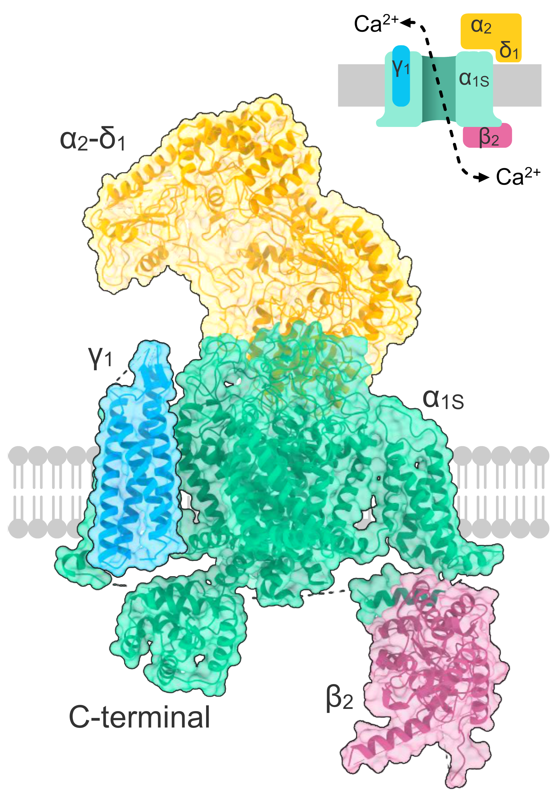
This little critter is found everywhere, including but not limited to: skeletal, cardiac and smooth muscle, the adrenal cortex and neurons throughout the central and peripheral nervous systems.
They do a bunch of other stuff too, but that's far beyond the scope of an FRCA exam-targeted post.
For now we'll focus on their cardiovascular handiwork, as this is what we're trying to counteract with our drugs.
Remind me how these channels work?
These are voltage-gated calcium channels that open slowly.
They do so at a membrane potential of around -30 mV, allowing calcium to flow into the cell, depolarising it further.
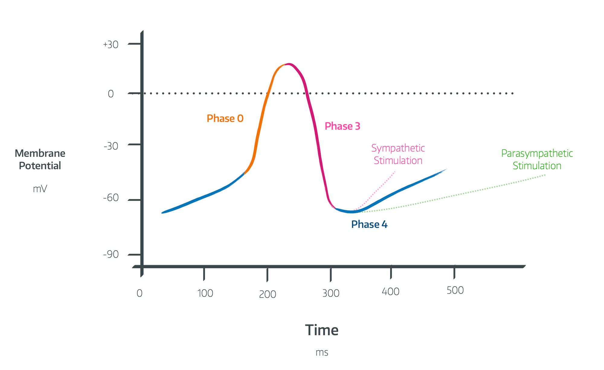
In the heart
In the pacemaker cells, this produces phase 0 of the pacemaker action potential
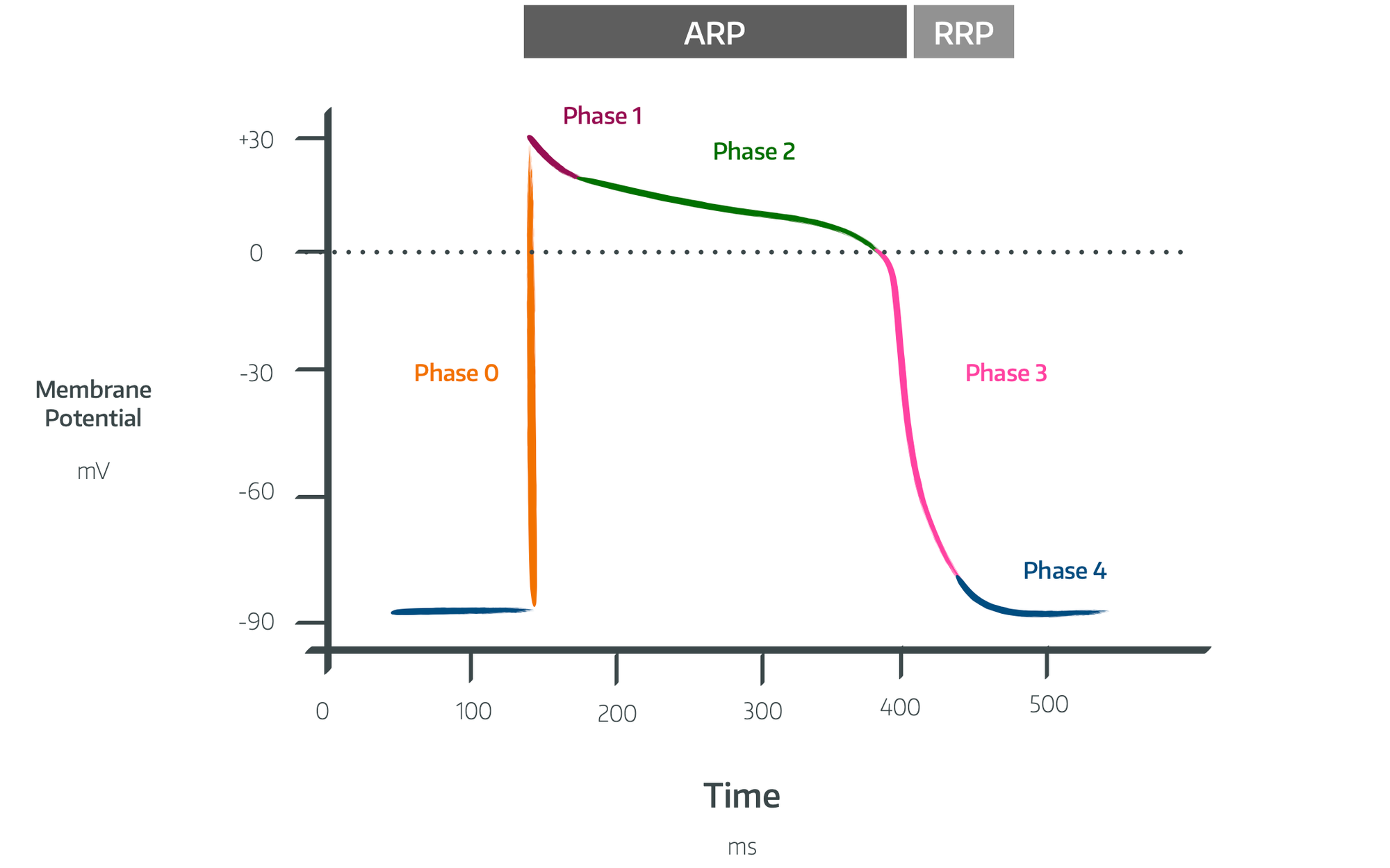
In the myocardial cells, this produces the phase 2 plateau of the cardiac action potential
In the blood vessels
In vascular smooth muscle, they modulate smooth muscle tone and facilitate contraction.
Remind me how smooth muscle contracts?
Well of course.
- An action potential causes membrane depolarisation and L-type calcium channels to open, allowing calcium influx
- This is complemented by further calcium release from the sarcoplasmic reticulum (calcium-induced-calcium release)
- Calcium binds calmodulin to form a complex
- This complex activates myosin light-chain kinase which phosphorylates myosin light chains
- This phosphorylated myosin can then interact with actin, causing smooth muscle contraction
- As intracellular calcium concentration falls, myosin light-chain phosphatase then dephosphorylates myosin, reduces the cross-bridge cycling, causing muscle relaxation